Sensory neurons shape γδ T cell effector programs to control Psoriasiform Inflammation.

Sensory neurons shape γδ T cell effector programs to control Psoriasiform Inflammation.
Inclan Rico, J.; Napuri, C.; Stephenson, A.; Rossi, H.; Femoe, U. M.; Musaigwa, F.; Hung, L.-Y.; Yu, H.; Luo, W.; Herbert, D.
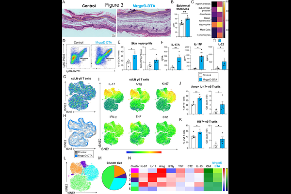
AbstractPsoriasis is a chronic autoimmune skin disorder marked by IL-17-producing {gamma}{delta} T cell ({gamma}{delta}T17) and pruritus, but immunoregulatory roles of itch-inducing neurons in this context remain unclear. This study addressed whether non-peptidergic (NP) afferents bearing the Mas-related G protein-coupled receptor D (MrgprD/NP1) and MrgprA3/NP2 subsets had differential effects on psoriasiform immunopathology. Data show human NP1 and NP2 neurons basally expressed an array of pattern recognition and cytokine receptor genes, and psoriatic human skin had a profound dysregulation of neuropeptides and their receptors. In mice, imiquimod (IMQ) application reduced the density of MrgprD+ skin afferents, whereas NP1 neuron ablation exacerbated IMQ-induced disease. Strikingly, NP1 activation using either optogenetics or {beta}-alanine before IMQ exposure significantly reduced epidermal thickness, psoriatic clinical score, and {gamma}{delta}T17 cell accumulation. In stark contrast, NP2 activation increased the numbers of {gamma}{delta}T17 cells that co-expressed amphiregulin (Areg) and exacerbated IMQ-driven skin pathology. Instead, pre-emptive NP1 stimulation shifted {gamma}{delta} T cell profiles away from being IL-17 and Areg dominant to IL-13+ {gamma}{delta} T cells expressing the transcription factor GATA3 accompanied by IL-10 secretion. Importantly, IL-10 signaling blockade reversed NP1-mediated suppression of IMQ-induced dermatitis. These data show that sensory neuron subsets can distinctly modulate inflammatory skin disease.