TCR-dependent and TCR-independent in-vitro T cell activation generate distinct functional, metabolic, and cytokine programs: Protein kinase C signalling augments anti-CD3+anti-CD28 responses

TCR-dependent and TCR-independent in-vitro T cell activation generate distinct functional, metabolic, and cytokine programs: Protein kinase C signalling augments anti-CD3+anti-CD28 responses
Ramteke, N. S.; Nandi, D.
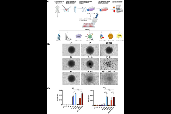
AbstractIntroduction: T cell activation is central to the adaptive immune response. In vitro studies on T cell activation often utilize two distinct approaches: first, engaging T cell receptors (TCR) using plate-bound anti-CD3 together with soluble anti-CD28 (TCR-dependent). Second, triggering intracellular signalling cascades using phorbol 12-myristate 13-acetate (PMA) and Ionomycin or P+I (TCR-independent). Both methods are widely used; however, a systematic comparison of the activation methods across a range of stimulation strengths to evaluate their effects on T cell function and metabolism has not been investigated in great detail. In this study, we compared the consequences of engaging T cells using TCR-dependent and TCR-independent activation pathways across varying signal strengths. Methods: T cells from BALB/c mice were isolated and activated under four conditions: anti-CD3, & anti-CD3+anti-CD28, PMA with low Ionomycin (P+IL) and PMA with high Ionomycin (P+IH). We studied differences with respect to several parameters: morphology, flow analysis, metabolic activities, cytokines. The roles of Protein kinase C (PKC) and Calcium; pathways were addressed by supplementing anti-CD3+anti-CD28 cultures with different doses of exogenous PMA or Ionomycin. Results: P+I activation outperformed the anti-CD3+anti-CD28 activation system across most readouts by displaying enhanced blasts, higher cycling, greater glucose uptake, increased lactate and ROS production, together with higher upregulation of CD25 and CD44 activation markers. P+IH activation dampened several responses including CD69 expression. CD4 co-receptor was downregulated greatly with P+I activation but not anti-CD3+anti-CD28. Most cytokines followed signal strength comparably between both systems; however, differences were observed with others: P+I stimulation favoured IL-6 and IL-12 induction whereas anti-CD3+anti-CD28 activation preferentially induced CCL2 and IL-1beta;. Importantly, PKC activity was substantially lower upon anti-CD3+anti-CD28 stimulation and the addition of PMA, but not Ionomycin, to anti-CD3+anti-CD28 cultures enhanced proliferation, metabolism and expression of activation markers. Discussion: TCR-dependent and TCR-independent T cell activation models have clear functional and metabolic differences. The observation that PKC signalling can boost T cell activation with anti-CD3+anti-CD28 is likely to be significant and may have translational implications such as CAR-T cell anti-tumor therapy where anti-CD3+anti-CD28 stimulation is widely used. The implications of our findings with regard to augmenting T cell mediated immunotherapies are discussed.