Cryo-electron tomographic analysis of anti-nephrin-mediated podocytopathy
Cryo-electron tomographic analysis of anti-nephrin-mediated podocytopathy
Birtasu, A. N.; Hengel, F. E.; Tomas, N. M.; Wieland, K.; Dehde, S.; Alves, J.; Scheffer, M. P.; Grahammer, F.; Huber, T. B.; Frangakis, A. S.
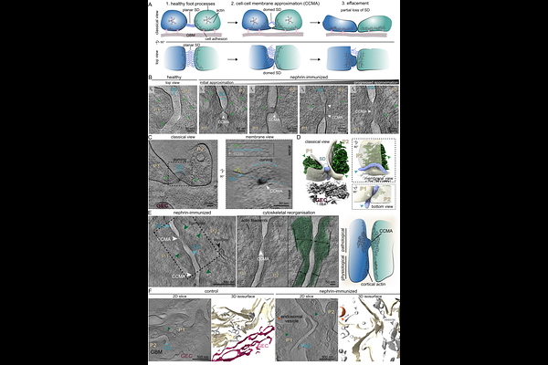
AbstractNephrotic kidney diseases are characterized by proteinuria caused by dysfunction of the glomerular filtration barrier. Here, we perform an ultrastructural analysis of anti-nephrin-mediated podocytopathy by visualizing the near-native slit diaphragm in situ across defined stages of disease progression using cryo-electron tomography. Our analysis shows that at early disease stages punctuate cell-cell membrane approximations appear, with basal membrane apposition between adjacent podocyte foot processes, while the initially planar, fishnet slit diaphragm progressively bends into a dome-shaped configuration towards the urinary space. At later stages, slit diaphragm disappearance coincides with the appearance of endosomal and vesicular structures and extensive cytoskeletal reorganization. These cryo-electron tomography-based observations provide a structural framework for understanding how antibody binding to nephrin translates into podocyte architectural failure.