Identification of loop regions as motifs determining cellular and organ chirality in Myosin 1C
Identification of loop regions as motifs determining cellular and organ chirality in Myosin 1C
Yamaguchi, A.; Sasamura, T.; Yoshimura, K.; Haraguchi, T.; Mori, T.; Ito, K.; Matsuno, K.
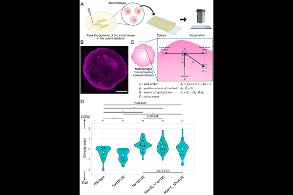
AbstractLeft-right (LR) asymmetry occurs throughout the animal kingdom, from microscopic to macroscopic scales; however, how microscale LR asymmetry (chirality) is integrated into higher-order structural LR asymmetry remains unclear. In Drosophila, the actin motor proteins Myosin1D and Myosin1C impose opposite chirality states on cell shape and intracellular F-actin flow, thereby directing dextral and sinistral morphogenesis, respectively. However, the protein motifs that confer their distinct activities in specifying chiral states have remained unknown. Here, we show that loop motifs within the myosin head domain that form actin-binding sites determine enantiomeric chirality. By swapping these loop motifs between Myosin1D and Myosin1C, we demonstrate that Myosin1D acquires sinistral activity when carrying Myosin1C loops. AlphaFold3 and molecular dynamics analyses reveal differences in loop structure and actin-binding energetics that may account for the contrasting chiral activities of Myosin1D and Myosin1C. Our findings identify specific protein motifs that dictate the handedness of actin flow and organ asymmetry, providing a mechanistic link between molecular interactions and macroscopic left-right patterning.