Microneedle delivery of stem cell derived retinal pigment epithelial cells
Microneedle delivery of stem cell derived retinal pigment epithelial cells
Ching, J.; Hinako, I.; Kitahata, S.; Morrow, L.; Kadonosono, K.
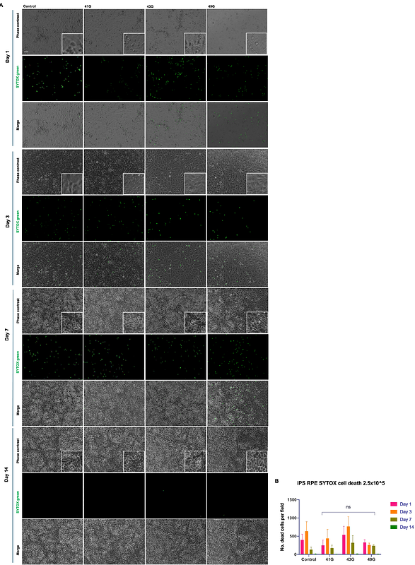
AbstractRetinal pigment epithelium (RPE) transplantation surgery has shown potential benefits in animal models and early clinical trials. However, current implantation techniques are limited by the need for large retinotomies when cell bearing scaffolds are used or cellular reflux in the case of cell suspension transplantation with micro-cannulae. Here we demonstrate that it is feasible to pass primary and stem cell derived RPE cells through microneedles with a bore as small as 30m. In vitro, there is no immediate loss of cell viability and this remains true throughout 14 days of live cell viability monitoring. Further, when the RPE cells are cultured over 14 days, they attach within 24 hours and become confluent, with normal morphological appearances. The formation of tight junctions is unimpeded with the typical polygonal pattern observed. The effects of shear stress due to passage of the cells through a small lumen were not evident in cultured cells labelled for the cytoskeletal protein F-actin. RPE cell VEGF secretion is unaffected by microneedle size in primary RPE and iPSC RPE cells. Ex vivo surgical implantation demonstrates that the technique is feasible, RPE cells engraft in the outer retina and we show that vitrectomy is not required to successfully transplant cells. Herein, we provide the first evidence of surgical microneedle cell delivery, which may have future applications more broadly within ophthalmic microsurgery and minimally invasive delivery strategies.