Quantitative 3D imaging of mouse and human intrahepatic bile ducts in homeostasis and liver injury
Quantitative 3D imaging of mouse and human intrahepatic bile ducts in homeostasis and liver injury
Hrncir, H. R.; Goodloe, B.; Bombin, S.; Zhang, Z.; Madabhushi, A.; Gracz, A. D.
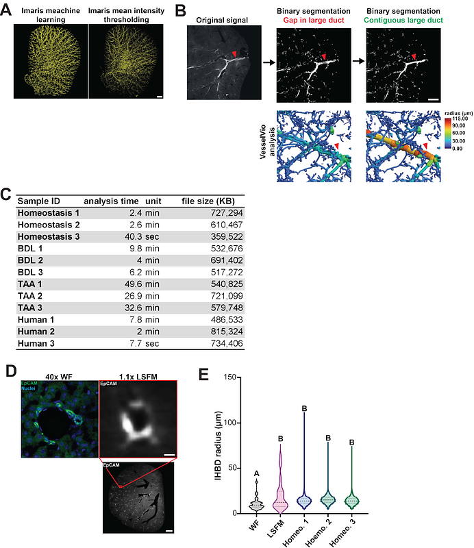
AbstractIntrahepatic bile ducts (IHBDs) form a complex hierarchical network essential for liver function. Remodeling and expansion of this network during ductular reaction (DR) is a hallmark of liver disease that can be a key indicator of disease severity. Conventional histology fails to capture the full extent of IHBD structural changes following injury due to the complex 3D organization of the IHBD network which limits understanding of DR, especially in human tissue. A major barrier to leveraging 3D imaging as a diagnostic tool is the absence of standardized pipelines for IHBD imaging and analysis. Here, we establish a robust 3D IHBD imaging and analysis workflow and apply it to both mouse and human liver tissues. This pipeline enables quantification of tissue and individual duct (\"segment\") level features and identifies features of invasive and noninvasive DR. In mouse models, we uncover regional phenotypes, including IHBD diverticula following duct blockage and the formation of anastomosed clusters after hepatocellular injury. Finally, we apply our 3D imaging and analysis workflow to quantify IHBD networks in human liver tissue. This work deepens our understanding of IHBD architecture in homeostasis and injury and lays the groundwork for advanced phenotyping of IHBD morphologies in mice and humans with relevance to next-generation experimental and diagnostic approaches to liver disease.