Mid-Infrared Photothermal Imaging of Fatty Acid Desaturation Reaction in Cancer Cells
Mid-Infrared Photothermal Imaging of Fatty Acid Desaturation Reaction in Cancer Cells
Teng, X.; Xia, T.; Yin, J.; Li, M.; Ao, J.; Ding, G.; Prabhu Dessai, C. V.; Isac, A. M.; Matei, D.; He, H.; Cheng, J.-X.
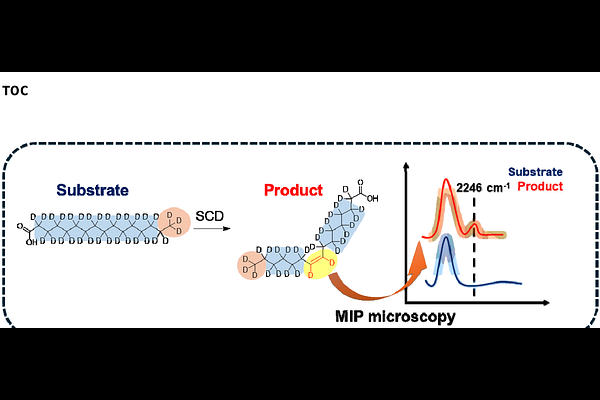
AbstractDirect visualization of metabolic conversions within living systems is essential for understanding metabolic activities yet challenging due to the absence of reaction-specific reporters and the limited sensitivity of current imaging modalities. Herein, we report an approach to monitor fatty acids (FAs) desaturation, primarily catalyzed by stearoyl-CoA desaturase, in cancer cells using deuterium (D)-labeled palmitic acid (PA-d31) as the reaction-specific reporter and mid-infrared photothermal (MIP) microscopy as the bond-selective imaging modality. The desaturation of PA-d31 produced a peak at 2246 cm-1 in the cell-silent region, corresponding to the stretching vibration of unsaturated C-D bonds (D-C=C-D) in unsaturated fatty acids. Penalized least squares fitting was employed to remove water background for enhancing the visibility of this peak. Our study revealed heterogeneous spatial distributions of both saturated FAs and their desaturated metabolites within lipid droplet pools in cancer cells. Furthermore, we observed an increase in fatty acid unsaturation level in OVCAR5 cells under cisplatin-induced stress. By directly visualizing fatty acid desaturation, this study offers new insights into fatty acid metabolism and opens avenues for evaluating new therapeutic strategies targeting fatty acid metabolism.