Visualizing phenotypic heterogeneity and single-cell morphology in situ during gut infection
Visualizing phenotypic heterogeneity and single-cell morphology in situ during gut infection
DiBenedetto, N. V.; Donnelly-Morell, M. L.; Kumamoto, C.; Shen, A.
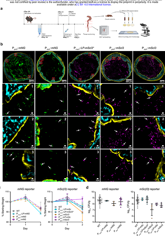
AbstractVisualizing phenotypic heterogeneity at single-cell resolution within dense microbial communities is technically challenging. Here, we present a method for visualizing this heterogeneity by combining spectrally compatible reporters to track the spatial distribution of gene expression in individual bacterial cells in the mammalian gut. Using toxin gene expression in Clostridioides difficile as a model for visualizing phenotypic heterogeneity, we demonstrate that, while C. difficile primarily occupies the lumen, a subpopulation of C. difficile associates with the colonic epithelium independent of toxin production. The approach further revealed that heterogeneity in C. difficile toxin gene expression is independent of location in the gut and unexpectedly showed that a toxin gene over-expressing mutant forms filamentous cells during the acute phase of infection. Thus, our reporter system provides quantitative, single-cell resolution of bacterial behavior within the intact gut environment and establishes a broadly applicable platform for investigating phenotypic heterogeneity in dense microbial communities.