Combination of 3D and 2D small and wide angle X-ray scattering imaging reveals diminished bone quality in the superior human femoral neck cortex
Combination of 3D and 2D small and wide angle X-ray scattering imaging reveals diminished bone quality in the superior human femoral neck cortex
Taenzer, T.; Kochetkova, T.; Baroni, A.; Simon, M.; Carlsen, M.; Zysset, P.; Bordin, S.; Guizar-Sicairos, M.; Liebi, M.
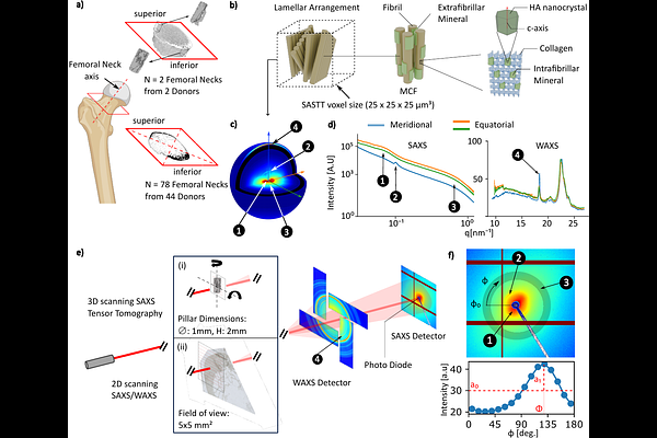
AbstractThe human femoral neck is particularly vulnerable to fracture, with failure most often initiating in the superior region. While age-related microstructural changes such as cortical thinning and increased porosity are well established, the contribution of material properties at the lamellar and mineralised collagen fibril (MCF) levels remains poorly understood. Here, regional differences in nanostructural properties of cortical bone from 78 femoral necks obtained from 44 donors aged 54-96 are investigated using a combined 2D and 3D X-ray scattering imaging approach. This approach quantifies MCF orientation and structure averaged over multiple lamellae in large fields of view, capturing tissue heterogeneity through the hierarchical scales. We identified misalignment between the scattering signals arising from the MCF bundles, specifically those associated with mineral inclusions in the collagen fibril gap regions, the mineral nanostructure, and the mineral crystal lattice, suggesting the presence of distinct mineral phases within and around the collagen fibers. Despite substantial intra-sample variability, the superior region displays on average more oblique MCF orientations, larger and thicker mineral platelets arranged in a less-ordered structure, greater misalignment between mineral and collagen at the MCF level, and possibly stiffer collagen fibres, with no significant trends observed with donor age or sex. The cumulative effect of these material property differences may contribute to the increased susceptibility of the superior cortex to compressive failure.