Boundary-guided cell alignment drives mouse epiblast maturation

Boundary-guided cell alignment drives mouse epiblast maturation
Ichikawa, T.; Guruciaga, P. C.; Hu, S.; Plunder, S.; Makino, M.; Hamaji, M.; Stokkermans, A.; Yoshida, S.; Erzberger, A.; Hiiragi, T.
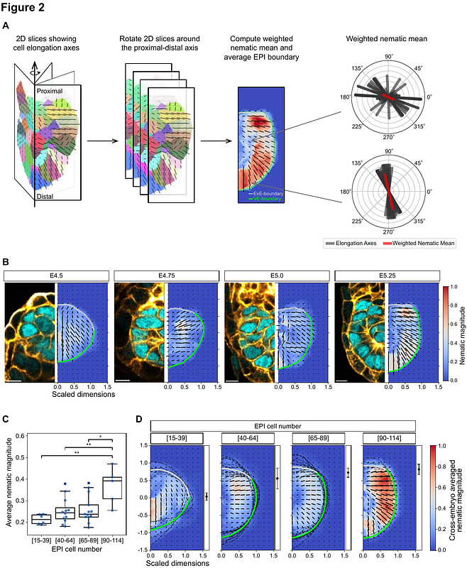
AbstractSymmetry breaking and pattern formation are critical events that occur throughout embryonic development. In early mouse development, a mass of non-polarized epiblast (EPI) cells in the blastocyst forms the egg-cylinder, while cells become apico-basally polarized and build a radial configuration. Yet, what drives the formation of this tissue architecture remains unclear. Here, we demonstrate that orientational patterning of EPI cells is dictated by heterogeneous tissue boundaries, which then defines central lumen positioning. We show EPI cells progressively orient perpendicular to the visceral endoderm (VE) boundary enriched with laminin and active integrin {beta}1, but parallel to the extraembryonic ectoderm interface. These orientation dynamics are consistent with general boundary-induced alignment effects in polar materials, with a topological defect predicting the position where the pro-amniotic cavity nucleates. Knockout of laminin {gamma}1 and integrin {beta}1 confirms the essential role of adhesion at the EPI-VE-boundary. The established EPI pattern, in turn, facilitates ERK activation to ensure proper EPI maturation. Together, these findings present the mechanistic basis and functional significance of EPI tissue patterning.