HIF-1α promotes osteogenic-angiogenic coupling response of BMSCs cell sheets
HIF-1α promotes osteogenic-angiogenic coupling response of BMSCs cell sheets
Zhang, D.; Teng, Y.; Huang, Y.; Liu, W.
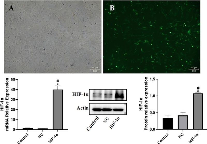
AbstractBackground: There is a critical need for management of vascularization in bone tissue engineering. The purpose of this study was to use HIF-1-transduced BMSCs fabricated prevascularized osteogenic cell sheets and explore HIF-1 promoted osteogenic-angiogenic coupling response of BMSCs cell sheets in vitro. Methods: HIF-1 was over-expressed by using a lentiviral vector, and transduced steadily in Wistar rats bone marrow mesenchymal stem cells (BMSCs). Real-time quantitative and western blot were performed to assess the expression level of HIF-1. Then, HIF-1/BMSCs were cultured to form osteogenic cell sheets(OCTs). ALP activity and Alizarin-red staining were performed respectively at day 14 and 21 to detect the characteristic of osteogenesis. Simultaneously, HIF-1/BMSCs were induced to differentiate into endothelial-like cells(iECs) for 14 days, and flow cytometry was further detected the conversion rates of HIF-1/BMSCs to iECs. Finally, iECs were seeded onto the OCTs to construct the prevascularized-osteogenic cell sheets (P-OCTs). In order to detect the role of HIF-1 involved in osteogenic-angiogenic coupling response of P-OCTs, Immunofluorescent staining for CD31 was performed on P-OCTs at 1, 3, 7, 14 days to check the formation of networks, and western blot of osteopontin(OPN) and osteocalcin (OCN) at 1, 7, 14 days to detect bone formation. Meanwhile, the none transduced BMSCs were as control. Conclusion: BMSCs were transduced by Lenti-HIF-1 with optimal multiplicity of infection was 30 (MOI=30), meanwhile, qPCR and western blot were performed to confirm this result. Next, the flow cytometric analysis results showed the conversion rate of BMSCs differentiated to iECs was 92.43%, which indicated BMSCs transduced by HIF-1 had a great superiority to differentiate into endothelial cells under experimental conditions. Then, ALP at day 14 and Alizarin-red staining at day 21 on OCTs showed an obvious osteogenic differentiation characteristic with more deep stained calcium nodules deposits than the control groups. Finally, we fabricated P-OCTs and observed iECs migrated reticulated fast and formed a large number of lumens and networks in experimental groups. At the same time, the results of Immunofluorescent staining for CD31 at day 1, 3, 7, 14 and osteogenic proteins expressions of OCN,OPN at day 1, 7, 14 showed that HIF-1 could promote osteogenic-angiogenic coupling response in P-OCTs significantly in vitro. All in all, the over-expressed HIF-1 of BMSCs cell sheets strategy can provide a new promising method for bone engineering, and we will detect further in vivo.